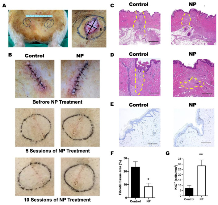
Figure 3.
Negative-pressure (NP) treatment in a porcine model. (A) Left: the design of the fusiform excision in the bilateral groin area to create wound tension. Right: the gross appearance of the wound after excision, which was fusiform and sized 2.5 cm in width and 3.5 cm in length. (B) The gross view of wound area before and after 5 and 10 sessions of negative-pressure treatment. (C) The granulation tissue (within the yellow dashed line) was observed in both the control and NP groups. (D) The magnification view of the granulation tissue in (C). Scale bar: 500 µm. (E) The proliferated keratinocytes are marked with Ki67 (brown). Scale bar: 200 µm. (F) The quantification of the fibrotic tissue area. (n = 4) (G) Quantification of Ki67+ cells per mm2 (n = 4). * p < 0.05, ** p < 0.01. Error bar: standard error.

