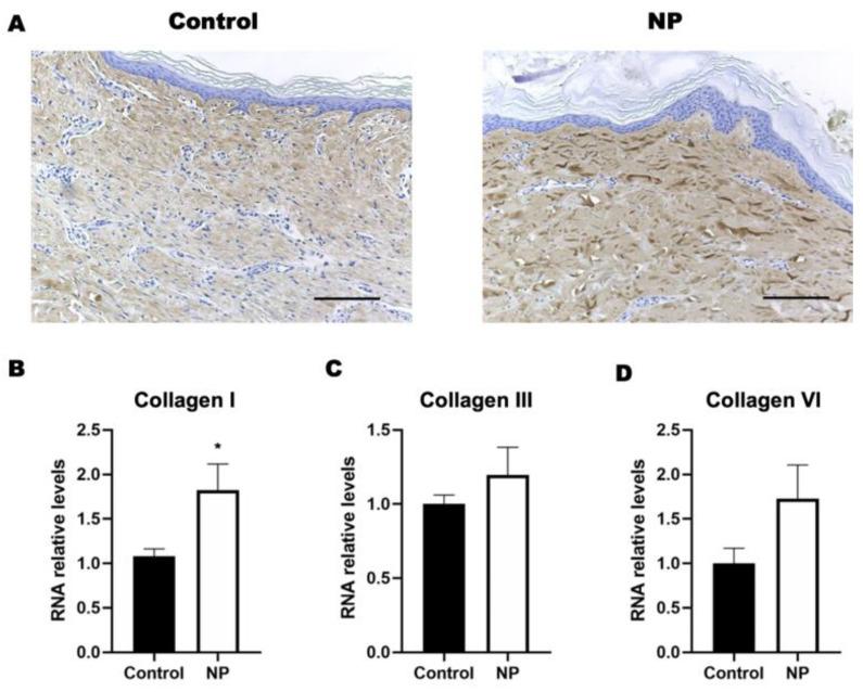
Figure 4.
The collagen expression in porcine skin tissue following negative-pressure treatment. (A) Histological staining of collagen type I in the control and NP groups. (B–D) The gene expression of collagens I, III, and VI in the control and NP groups (n = 4). Scale bar: 200 µm. * p < 0.05. Error bar: standard error.

