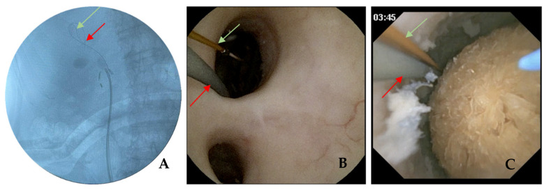
Figure 2.
Pressure wire placement intro renal pelvis: (A) fluoroscopic image of PressureWire (green) and safety wire (red) in the renal pelvis. (B) Endoscopic vision of the renal pelvis with a PressureWire (green) and safety wire (red) going inside the upper calyx. (C) Endoscopic vision before starting lithotripsy of a dihydrate calcium oxalate stone with a safety wire (red), PressureWire (green) and fibre laser in the renal pelvis.

