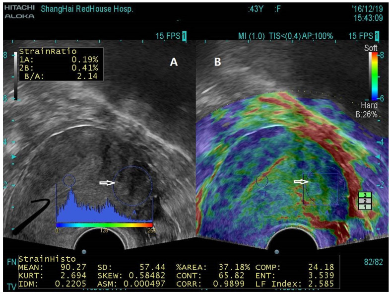
Figure 2.
Transvaginal ultrasound image for a patient with a small uterus (59 × 58 × 57 mm) who complained of moderate dysmenorrhea with elevated CA125 level and was suspected with AM. (A) The big, circled area (white arrows) showed the ROI. The conventional B-mode TVUS image showed no sign that was consistent with a typical or spherical enlarged uterus or the presence of mild but not severe or obvious internal inhomogeneous echo in ROI. (B) Transvaginal elastosonography image showing an increased stiffness value (LFI = 2.585) in the same ROI shown in the TVUS (white arrow), indicative of adenomyosis. AM indicates adenomyosis; LFI, liver function index; ROI, region of interest; TVUS, transvaginal ultrasound. Replicated from Liu et al. [71] (Reprinted with permission from Reproductive Sciences).

