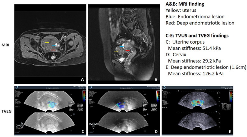
Figure 3.
The use of ultrasonic elastography to diagnose deep endometriosis and its consistency with MRI findings. For this patient who complained of severe dysmenorrhea, both MRI (A,B) and a shear-wave ultrasonic elastography (C–E) were used to diagnose deep endometriosis. While the anatomical localization of the deep endometriotic lesion is not as straightforward as MRI, the elastography gave a lesional stiffness value, and in this case the lesion is quite stiff and thus highly fibrotic. Note that for this shear-wave elastography which gives out absolute tissue stiffness value in kilo Pascal (kPa), the color key is reversed, with the blue color depicting the softest while the red color indicating the hardest tissues. More detailed explanation for each figure is given at the upper right panel. (Courtesy of Dr. Ding Ding).

