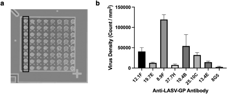
Figure 4.
Screening of anti-LASV GP antibodies on SP-IRIS. (a) Image of the SP-IRIS chip spotted with eight of the Lassa antibodies. Each column, shown by the black rectangle, represents one antibody type. The rest of the antibodies were spotted on a different chip. Spots have a diameter of approximately 150 μm. (b) Average virus densities captured on each antibody for 8 replicate spots following an incubation with 107 PFU/mL rVSV-LASV for 1 h. Wild-type VSV-specific 8G5 antibody was also spotted on the chip to obtain the detection threshold, which is calculated as 7700 particles/mm2. Only the antibodies that had a signal over the detection threshold and negative antibody are shown in the bar graph for the sake of simplicity. Results given in Fig. 4 are representative of at least two independent experiments.

