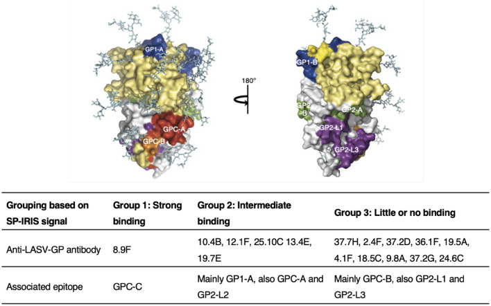
Figure 6.
LASV antibody epitope mapping based on SP-IRIS data. Top image shows the surface view of LASV GP monomer structure. GP1 is colored yellow and GP2 is colored white. Epitope color code: GP1-A and GP1-B, blue; GP2-A and GP2-B, green; GP2-L1-3, purple; GPC-A, red; and GPC-B, orange. The location of the conformational epitope of GPC-C, where 8.9F binds, is unknown. Reproduced with permission from 49. Bottom table groups the LASV antibodies based on their binding levels on SP-IRIS. The only Group 1 antibody 8.9F binds to a unique region in GPC-C and it is the only antibody that showed a strong binding on SP-IRIS. Group 2 antibodies show intermediate binding on SP-IRIS and mainly binds to the GP1 region of the glycoprotein. Group 3 antibodies, which show little or no binding on SP-IRIS, mostly binds to GPC-B and also GP2-L1 and GP2-L3 linear regions.

