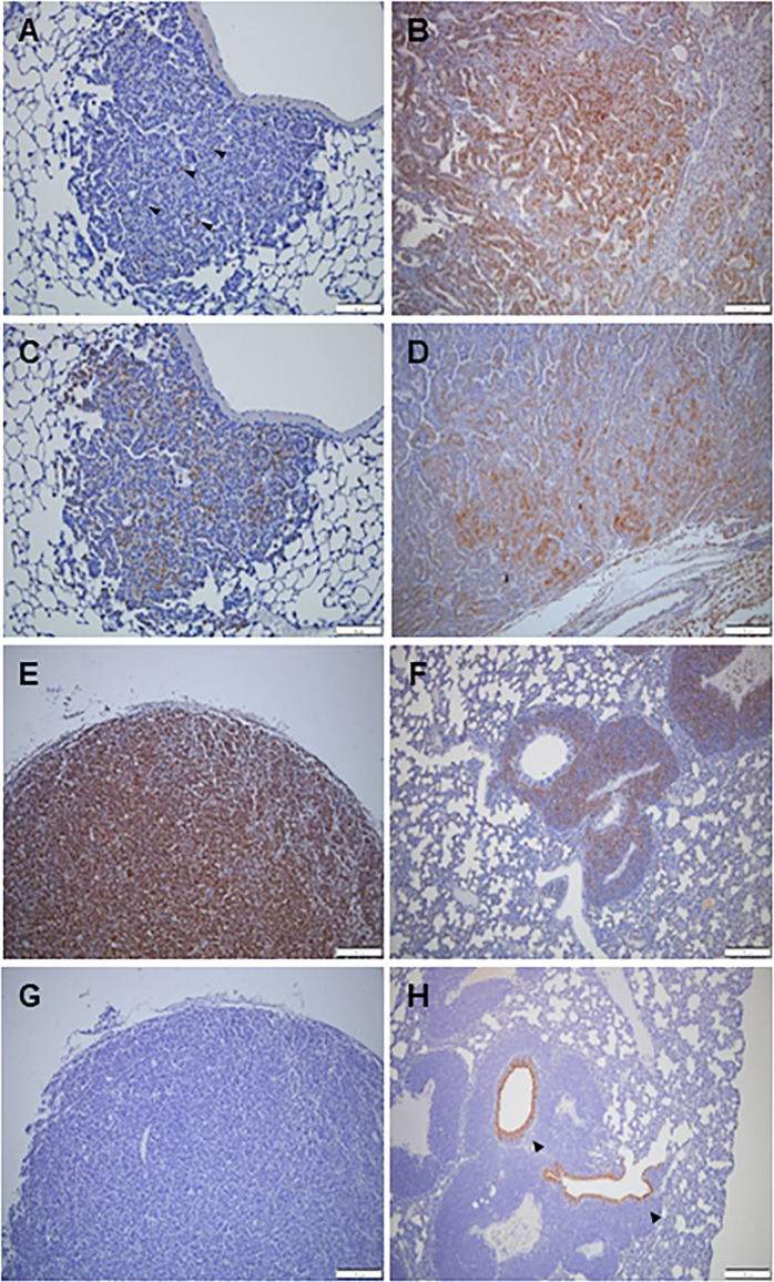
Fig 6. Representative images of immunohistochemical staining.
A few PCNA-positive cells are shown in adenoma of the lung (A) Many PCNA-positive cells were found in lung adenocarcinoma (B), thymic malignant lymphoma (E), and thymic malignant lymphoma (TML) metastasis in the lungs (F). A few SPC-positive cells indicate an adenoma of the lungs (C), but many SPC-positive cells indicate adenocarcinoma of the lung (D). No CC10-positive cells were observed in the adenoma of the lungs (G), but CC10-positive cells were present in the bronchial epithelium at the center of the TML lung metastasis. Scale bar represents 100 μm.

