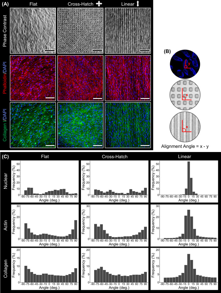
Figure 2.
Characterization of cell and ECM alignment of DPC sheets cultured on flat or microgrooved substrates. (A) Phase contrast images, phalloidin staining (red), and collagen I (green) immunostaining of the DPC sheets show that the DPCs cultured on the linear microgrooves aligned in parallel with the linear microgrooves and generated an aligned ECM. (B) Schematic of the alignment quantification method. (C) Nuclear, actin cytoskeleton, and collagen I alignment were quantified for the cell sheets cultured on different topographies, validating that these features were linearly oriented on the linear but not on the flat or cross-hatch substrates. Scale bar: (A) 100 μm.

