Figure 3.
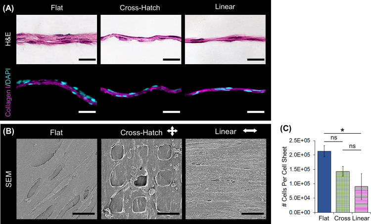
Structural characterization of the DPC sheets. (A) H&E staining illustrates that sheets cultured on all topographies were solid and cellular and those formed on the microtopographies were less thick than those cultured on the flat substrate. Immunostaining verifies that the DPC sheet ECM comprises type I collagen (magenta); nuclei were stained with DAPI (blue). (B) SEM further confirmed that the cell sheets contained a continuous matrix and that the sheets aligned their matrix when linear topographies. (C) Cell sheets cultured on the flat substrate contained more cells than those cultured on microtopographies (*: p-value < 0.05). Scale bars: (A) histology = 50 μm; (B) SEM = 15 μm.

