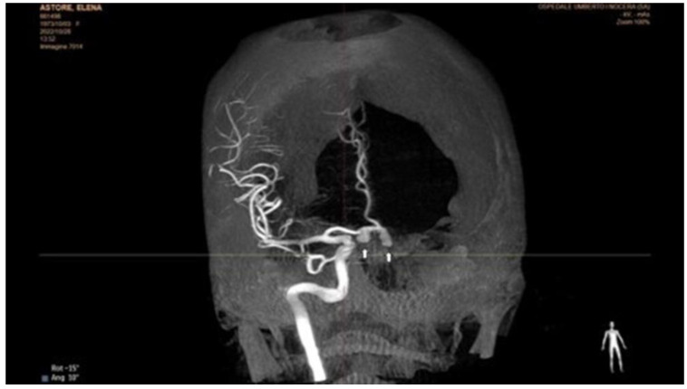
Figure 2.
Selective rotational angiography of the right internal carotid artery. The exam shows two aneurysmal dilatations, one of which is sac-shaped in the portion of the right midsection, and the second in correspondence with the ipsilateral A1–A2 angle. Both have the bottom of the aneurysm directed downwards. The first extension has a large collar, and the second of 1–2 mm.

