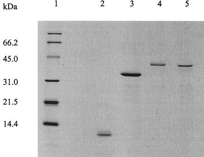
FIG. 1.
SDS-PAGE analysis of purified recombinant M. tuberculosis antigens. One microgram of protein was loaded in each lane. Lane 1, molecular weight standard; lane 2, recombinant ESAT-6; lane 3, recombinant Ag85B; lane 4, Ag85B-ESAT-6 fusion protein; lane 5, ESAT-6-Ag85B fusion protein. Protein bands were visualized by Coomassie blue staining.

