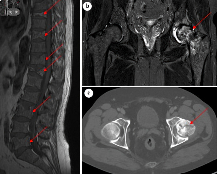
Figure 1.
Sagittal T1-weighted MRI of lumbar and thoracic spine (a) showing multiple vertebral fractures and osteopenia (arrows). Coronal STIR MRI (b) and axial CT image (c) of the hip joints with a suspected malignant process in the left proximal femur and a subcapital fracture (arrows), with accompanying synovitis and accumulation of fluid in the left hip joint. MRI: magnetic resonance imaging; CT: computed tomography.

