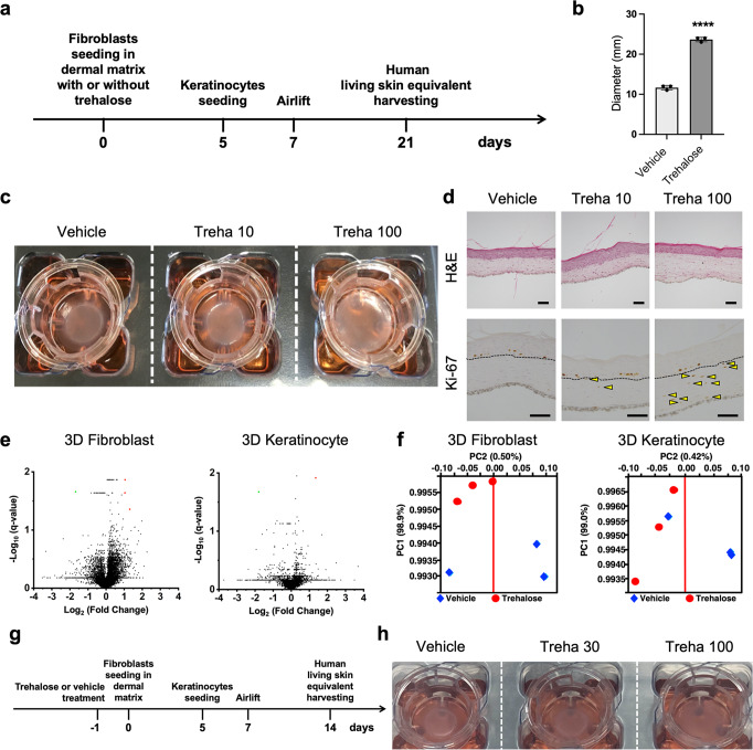
Fig. 1. Effect of trehalose in the preparation of living skin equivalents.
a Schematic for the preparation of cultured skin equivalents. b Diameters of LSEs with or without trehalose (100 mg/ml) added in the collagen gel, prepared in the Transwell-COL with 24-mm insert in a six-well culture plate after 2-week airlifting at 37 °C. Data were expressed as means ± SD for three LSEs, which are representative of three independent experiments with similar results. ****P < 0.0001 versus vehicle control groups using the Student t-test. c Macroscopic pictures of LSEs with or without trehalose (10 and 100 mg/ml) added in the collagen gel after 2-week airlifting. d LSEs stained with hematoxylin and eosin (Scale bar = 50 μm). Paraffin-embedded sections of LSEs were sectioned and subjected to immunohistochemistry with Ki67 antibody. Yellow arrowheads indicate the Ki67 positive fibroblasts in the dermis (Dotted line: dermal–epidermal junction, Scale bar = 100 μm). e Volcano plots showing gene expression in the absence or the presence of trehalose. Red or green rounds indicate genes that increased by more than 2-fold or decreased by less than half, respectively, with less than 0.05 of q values. f A principal component analysis (PCA) with gene expressions in the absence or the presence of trehalose showed no clear separation between principal component PC1 and PC2. g Schematic for the preparation of cultured skin equivalents with fibroblasts treated with or without trehalose before seeding in the dermal matrix. h Representative picture of LSEs with the fibroblast treated with or without trehalose (30 and 100 mg/ml) before seeding in the collagen gel after 1-week airlifting at 37 °C. Data were representative of three independent experiments.

