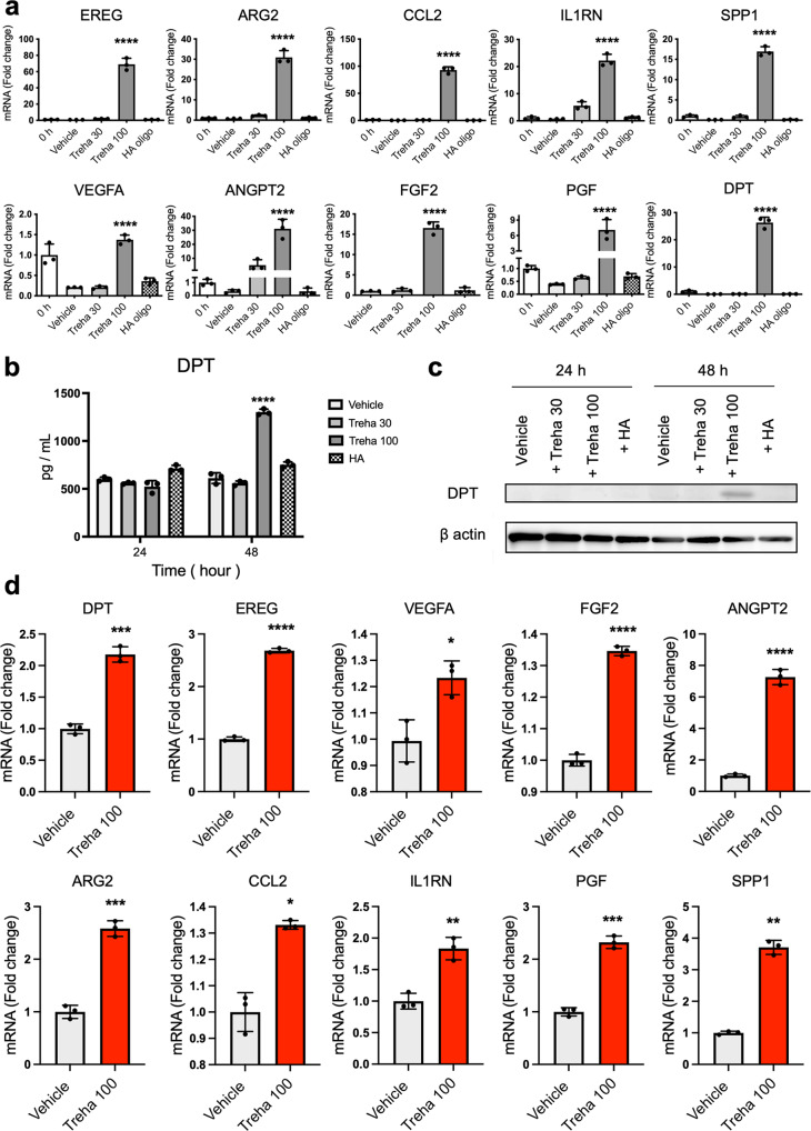
Fig. 6. Trehalose induces an increase in the expression of wound healing-related molecules.
a Human dermal fibroblasts were treated with trehalose (30 and 100 mg/ml), tetrasaccharide hyaluronan (HA oligo) (30 μg/ml), or vehicle control (PBS) for 24 h. EREG, ARG2, CCL2, IL-1RN, PGF, SPP1, VEGF, ANGPT2, and DPT mRNA expressions were assessed by qPCR. Data were shown as relative expression to the control (0 h) fibroblasts. For FGF2 mRNA expression, data are shown as relative expression to vehicle control groups at 24 h. b DPT was measured by ELISA in the culture medium of human dermal fibroblasts. One set of fibroblasts was treated with trehalose 30 or 100 mg/ml, tetrasaccharide hyaluronan (HA) (30 μg/ml), or the vehicle (PBS) for 24 or 48 h (n = 3). c Representative Western blots showing DPT and β-actin expression in human dermal fibroblasts 24 or 48 h after vehicle, trehalose 30 or 100 mg/ml, tetrasaccharide hyaluronan (HA) (30 μg/ml) exposure. d Trehalose (100 mg/ml) or vehicle were added in the human dermal fibroblasts populated collagen gel for 72 h. DPT, EREG, VEGF, FGF2, ANGPT2, ARG2, CCL2, IL-1RN, PGF, and SPP1 mRNA expression were assessed by qPCR. Data were shown as the relative expression to the control (vehicle-treated). *P < 0.05, **P < 0.01, ***P < 0.001, ****P < 0.0001 versus the vehicle-treated control group by one-way ANOVA (a, b) or Student t-test (d). Data were expressed as means ± SD for three wells (a, b) or three dermal substitutes (d), and representative of three independent experiments.

