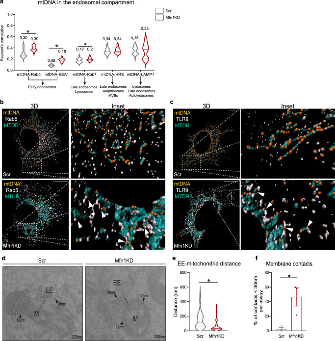
Fig. 4. Mfn1 deficiency causes preferential location of mtDNA and mitochondria in early endosomes and promotes close contacts between Rab5+ early endosomes and mitochondria.
a Quantification of Pearson’s correlation between mtDNA and endosomal markers (n = 20 images per condition, except for mtDNA-LAMP1, where n = 39). Representative 3D reconstructions of immunostainings using dsDNA with nuclear subtraction (mtDNA, orange), mitochondria with Mitotracker Deep Red (turquoise) and b Rab5 (white) or c TLR9 (white) (Scale bar,10 µm). d Representative images of immunogold staining of Rab5 (gold particle 18 nm) and SdhA (gold particle 12 nm) in Scr- and Mfn1KD myoblasts (Scale bar, 200 nm). e Quantification of the distance between marked early endosomes and mitochondria in Scr (n = 38 contacts) and Mfn1KD myoblasts (n = 31 contacts). f Percentage of measured contacts <30 nm in Scr- and Mfn1-deficient muscle cells (n = 3, each point represents the mean of quantifications obtained in three independent experiments). b, c Arrows point to positive co-distribution. d Arrows point gold particles. a, e, f Two-sided Students’ t test. Data are expressed as the mean of n independent experiments ± SEM.*p vs. Scr <0.05. a, e, f Source data is provided in the Source Data File.

