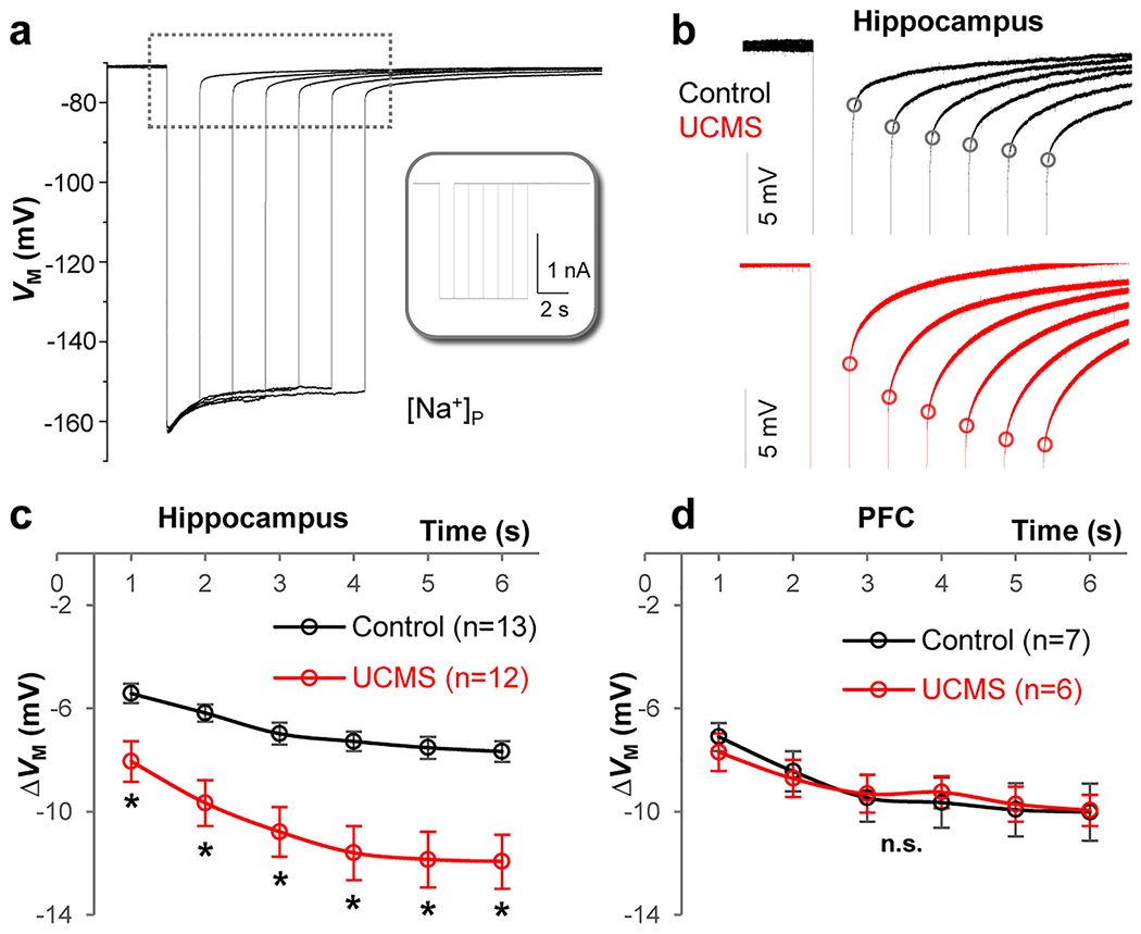
Figure 5: UCMS impairs the K+ redistribution capacity of an astrocyte syncytium.

(a) Representative trace recorded with K+ free-Na+ containing electrode [Na+]P in current-clamp mode. Inset: −2 nA current steps (Iholding) were applied at incremental durations from 1 to 6 s. In between these steps, the cell was maintained at resting condition for VM recovery. The longer the duration of the current steps, the stronger the negative shift in the reversal potential (Vrev, black dots) upon withdrawal of the steps, indicating more accumulation of K+ inside astrocytes. (b) Enlarged recording trace (indicated by dashed line area in a) of astrocytes in the hippocampus from control (black) and UCMS (red) animals. The ΔVM is the difference between the basal VM and Vrev values and is used to compare the capacity of K+ redistribution in different groups. (c) In the hippocampus, the increased ΔVM in the UCMS group indicates weakened capacity of K+ redistribution of the astrocyte syncytium. (d) In the PFC, there was no significant difference of ΔVM between control and UCMS groups. Two-Way Mixed-Design ANOVA. *: p < 0.05. n.s.: not significant. n = 6-13 recorded cells per group.
