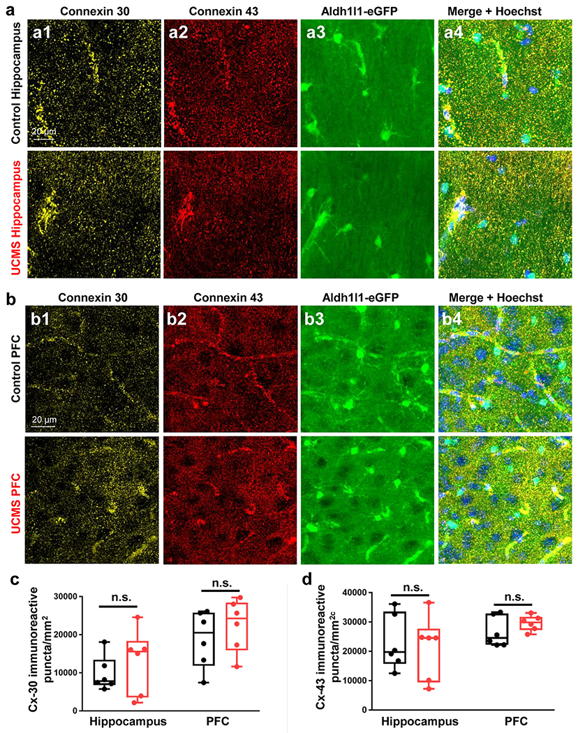
Figure 6: UCMS does not alter the number of connexin 30 (Cx30) or connexin 43 (Cx43)-immunoreactive puncta in the hippocampus or PFC.

(a1-a4) Representative 40X immunofluorescent images of connexin 30 (yellow), connexin 43 (red), Aldh1l1-eGFP transgene (green), and Hoechst (blue) in the stratum radiatum of the hippocampus of control (top row) and UCMS exposed (bottom row) animals. (b1-b4) Representative 40X immunofluorescent images of connexin 30 (yellow), connexin 43 (red), Aldh1l1-eGFP transgene (green), and Hoechst (blue) in the PFC of control (top row) and UCMS exposed (bottom row) animals. (c) Graphical representation of the number of connexin 30 immunoreactive puncta per area of hippocampus/PFC in control and UCMS mice. (d) Graphical representation of the number of connexin 43 immunoreactive puncta per area of hippocampus/PFC in control and UCMS mice. Data were analyzed using Student’s t-test. n.s.: not significant. N = 6 animals per condition.
