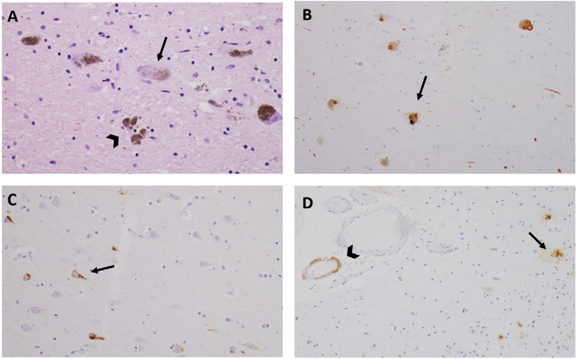
FIGURE 3.

Histopathological findings for a patient with mixed Lewy body dementia and Alzheimer disease neuropathologya
aMicroscopic views of the midbrain substantia nigra with hematoxylin and eosin (panel A) and synuclein immunohistochemistry (panel B) demonstrate Lewy bodies (arrows) and extraneuronal pigment (arrow-head). Tau immunohistochemistry on Ammon’s horn of the hippocampus indicates the presence of neurofibrillary tangles (panel C). β-amyloid immunohistochemistry of the frontal cortex indicates the presence of β-amyloid plaques (arrow) and cerebral amyloid angiopathy (arrowhead) (panel D).
