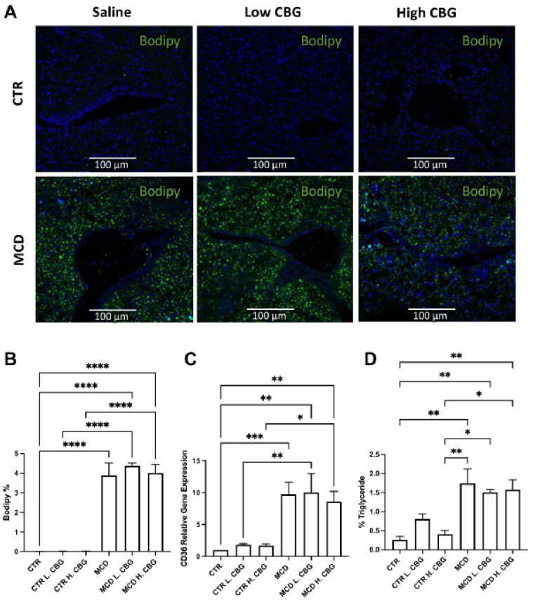
Figure 2.
Steatosis was evaluated using histology and RT-qPCR. Immunofluorescence staining of bodipy (green) and DAPI (blue) (A, 20×). Immunofluorescence staining was quantified using ImageJ (B). mRNA expression of CD36 (C) was measured via RT-qPCR, and the percentage of triglyceride was evaluated using a commercially available ELISA kit (D). * p < 0.05, ** p < 0.01, *** p < 0.001, **** p < 0.0001.

