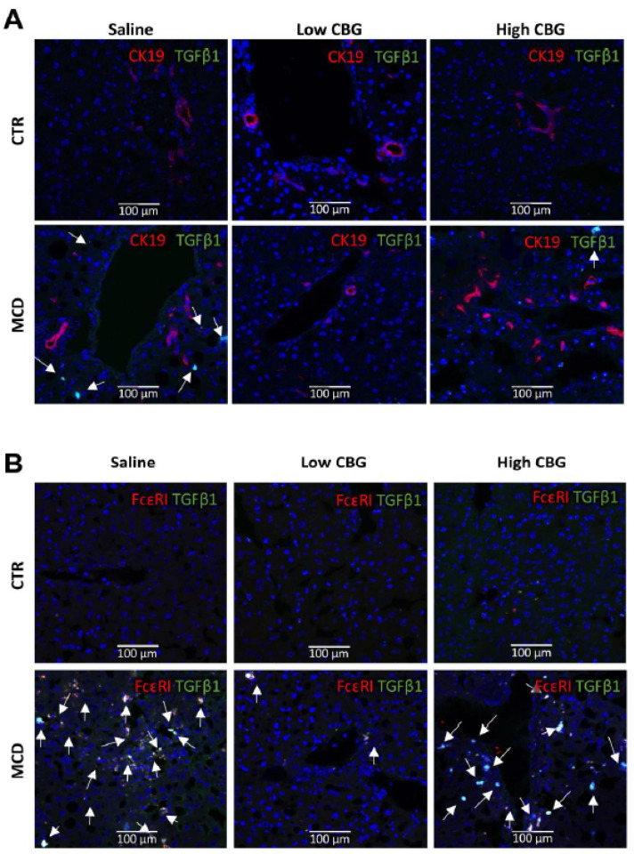
Figure 6.
Colocalization of the proinflammatory marker TGF-β1 with the cholangiocytes (CK19+) or mast cells (FcεR1+) in mice liver tissue. (A) Immunofluorescence co-staining of cholangiocytes biomarker CK19 (red) and proinflammatory factor TGF-β1 (green), and DAPI (nuclei stained in blue). CK19+ cells were not co-localized with TGF-β1 (white arrows). (B) immunofluorescence co-staining of mast cell biomarker FcεR1 (red) and proinflammatory factor TGF-β1 (green), and nuclei (blue). Strong colocalization of TGF-β1 with mast cells in MCD groups (white arrows). Pictures are taken at 20× magnification.

