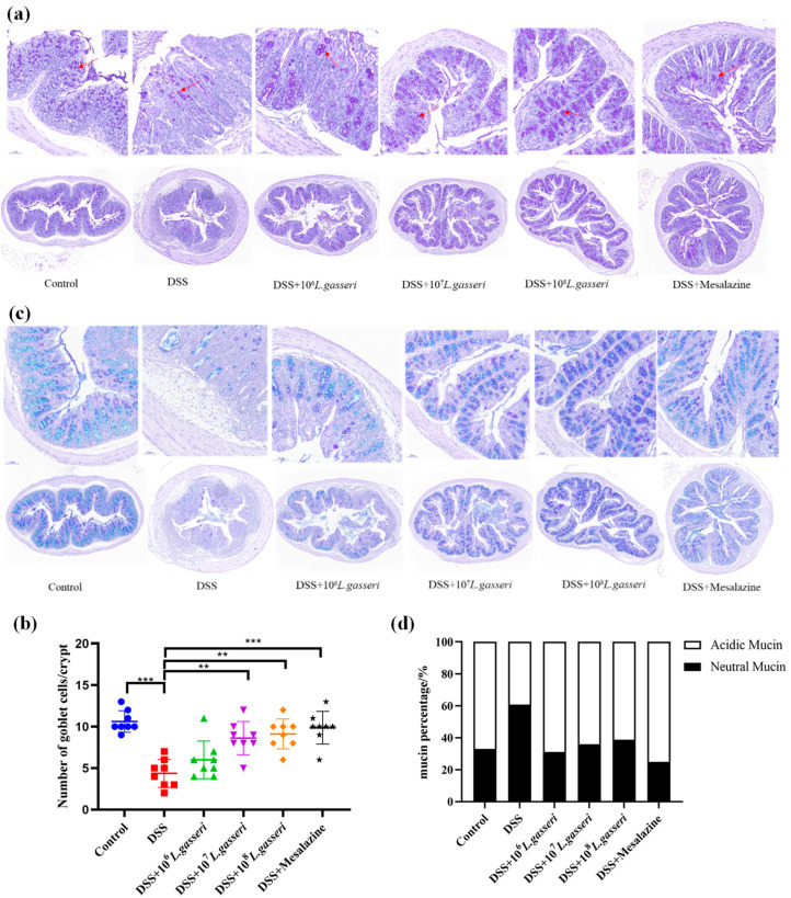
Figure 6.
Staining and distribution of goblet cells and mucin. (a) Staining of goblet cells in colonic tissue, (b) counting of goblet cells in colonic tissue, (c) staining of mucin and distribution in colonic tissue, and (d) distribution of mucin in colonic tissue. ** p < 0.01, *** p < 0.001 represent significant difference between groups.

