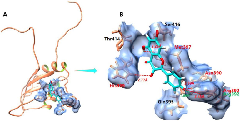
Figure 7.
(A,B) shows the kaempferol–EWS complex. (A) illustrates the global structure of the complex, and (B) focuses on the binding pocket. The salt bridges and hydrogen bonds formed in the docked complex are shown in green and red color, respectively. The EWS protein is colored in coral, and helix interiors are colored in chartreuse green while the surface of the binding pocket is colored in light blue.

