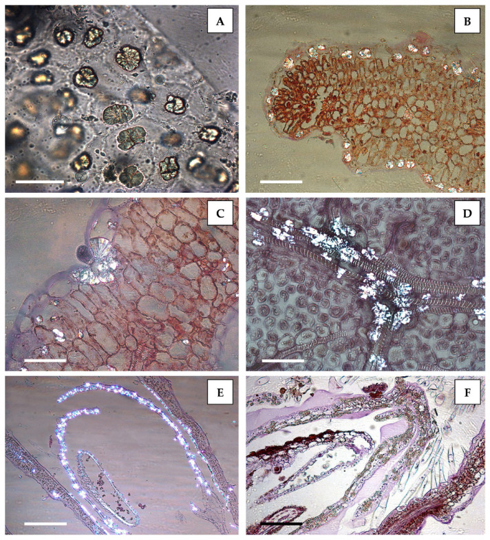
Figure 1.
Spherocrystalline masses of diosmin were abundant in the leaf epidermal cells (A,B), sometimes crowded at the base of trichomes (C), as well as along the leaf veins (D). These crystals were also visible inside the thin petals of the flowers (E,F). A: Leaf peelings mounted in water, where diosmin spherocrystals appear unstained. (B–F): Paraffin-embedded samples and semithin sections stained with hematoxylin–eosin. Cross section (B,C) and transdermal section (D) of a leaf; longitudinal section of a flower (E,F). Aggregates of diosmin crystals were birefringent under polarized light in both leaves and flowers (B–E) but remained unstained when treated with hematoxylin–eosin staining (F). Bars: A = 50 µm; B = 100 µm; C = 50 µm; D = 50 µm; E = 100 µm; F = 100 µm.

