Figure 1.
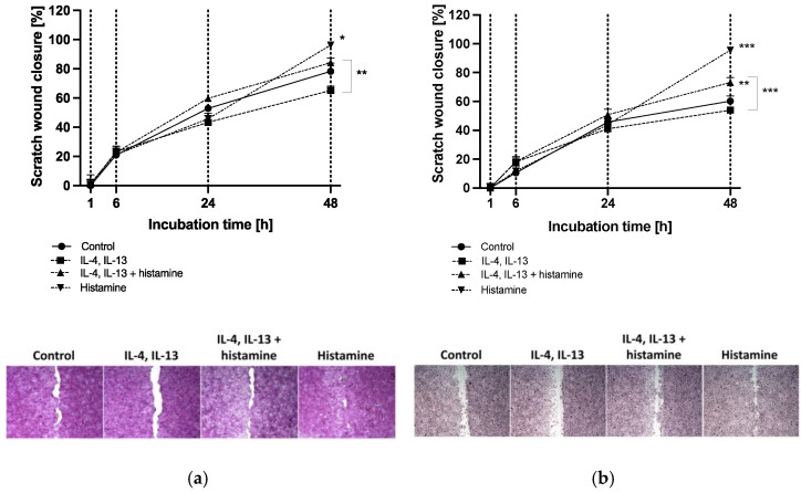
(a) HaCaT keratinocyte and (b) primary keratinocyte scratch wound closure after 1, 6, 24 and 48 h of incubation. Keratinocytes were either stimulated with TH2 cytokines (50 ng/mL IL-4, 50 ng/mL IL-13) with or without 10 µM histamine or cultivated under normal medium conditions with 10 µM histamine. The control group was cultivated without any stimulation. Wound closure is displayed as scratch closure and is presented as mean ± standard error of the mean (SEM) in [%]. Two independent experiments were performed as replicates. Scratch assays were evaluated using six images per sample. Asterisks [*] indicate significant deviations from the control at the respective time point (* p < 0.05, ** p < 0.01 and *** p < 0.001). Cells were stained with haematoxylin-eosin. Images show scratch wound closure of (a) HaCaT keratinocytes and (b) primary keratinocytes after 48 h of incubation.

