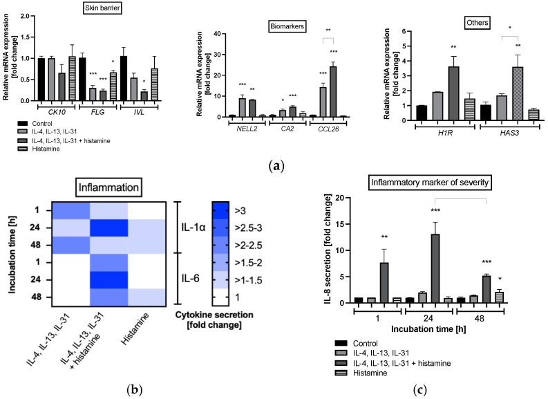
Figure 2.
(a) Gene expression profiles of primary keratinocytes after 48 h of incubation and (b) interleukin (IL) IL-1α, IL-6, (c) IL-8 protein expression at after 1, 24 and 48 h of incubation. Keratinocytes were stimulated with TH2 cytokines (50 ng/mL IL-4, 50 ng/mL IL-13, 25 ng/mL IL-31) either with or without 10 µM histamine or under normal medium conditions with 10 µM histamine in the absence of TH2 cytokines. The control group was cultivated under normal medium conditions without any stimulation. (a) Transcript levels are given as normalized relative mRNA expression compared to the untreated control. (b,c) Cytokine levels are given as fold changes compared to the untreated control at the respective time point. (a,c) All data are presented as mean ± SEM [fold change]. (b) All data in the heatmap are presented as mean [fold change]. Two independent experiments were performed as replicates. Asterisks [*] indicate significant deviations from the untreated control (* p < 0.05, ** p < 0.01 and *** p < 0.001).

