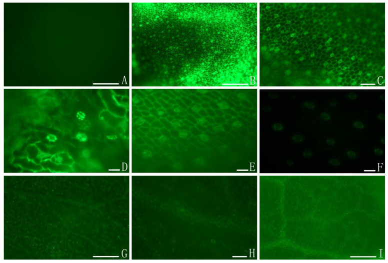
Figure 2.
The colonization of I. cateinannulata submerged conidia in buckwheat leaf as assessed via fluorescence microscopy. Note: (A) In control samples, no fluorescent aggregation was observed, consistent with an absence of submerged conidia colonization. (B–D) At 0.5 to 1 h after spraying, significant fluorescent signal was visible and distributed around veins and stomata. (E,F) At 2 to 8 h after spraying, the fluorescence intensity of veins and stomata decreased. (G–I) At 4 to 12 h after spraying, fluorescence was no longer visibly associated with veins or stomata, and the leaves presented a uniform color brighter than that of control leaves (I). Some bright spots were visible on the blade surface (G,H), which may correspond to small amounts of conidia attached to the blade surface. Scale bar: 1 mm.

