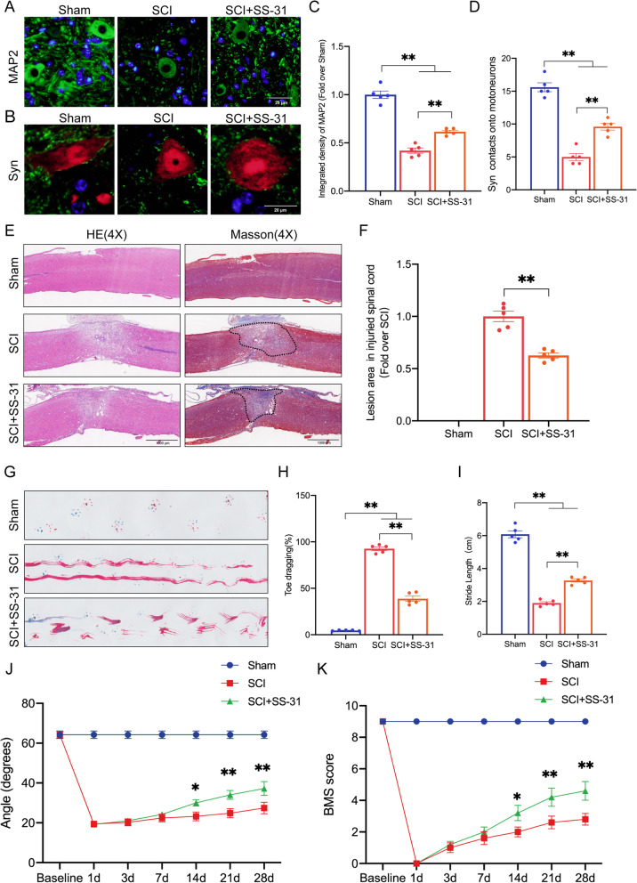
Fig. 1.
SS-31 facilitates functional recovery following SCI. A Photographs of spinal cord sections in the respective groups stained with antibody MAP2 (green) (scale bar = 25 μm). B Photographs of spinal cord sections in the respective groups stained with Syn (green)/NeuN (red) (scale bar = 20 μm). C MAP2 optical density within a spinal cord subjected to injury on day 28. D Relevant quantitative results for motor neuron-contacting synapse amounts on day 28 after SCI. E Longitudinal spinal cord sections from the groups at 28 dpi were examined via HE dyeing and Masson dyeing (scale bar = 1000 μm). F Quantitative investigations of Masson-positive lesions within the spinal cords of the respective groups. G Photographs of mouse footprints on day 28 following SCI. Blue: fore paw print; Red: hind paw print. H, I Toe dragging (%) and stride length (cm) analyses of mice at 28 dpi. J, K Inclined plane test and BMS results for the indicated groups at days 0, 1, 3, 7, 14, 21, and 28. The data are shown as the mean ± SEM. n = 5. *P < 0.05, **P < 0.01. ns indicates no significance

