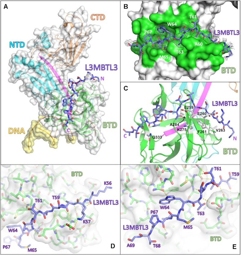
Figure 3.

RBPJ–L3MBTL3–DNA crystal structure. (A) The RBPJ–L3MBTL3–DNA X-ray structure with the NTD, BTD and CTD colored cyan, green and orange, respectively. The DNA wire model is shown in yellow. L3MBTL3 RBP-ID (55–70) represented as purple sticks binds as an elongated peptide along the top and front faces of the BTD. (B) Figure shows the L3MBTL3 binding pocket (colored green) on the BTD of RBPJ. RBPJ residues that directly contact L3MBTL3 were determined by the PISA server(41). The 2Fo– Fc electron density map contoured at 1σ corresponds to the L3MBTL3 peptide. (C) L3MBTL3 binds residues in the BTD, depicted as grey sticks, that are important for binding RAM and other RAM-like coregulators. (D, E) Panels show polar interaction network (yellow dashed lines) between RBPJ and the N-terminal (D) and C-terminal (E) regions of L3MBTL3.
