
FIGURE 1.
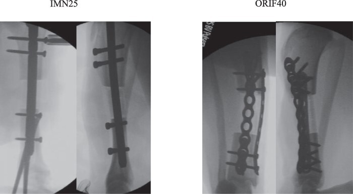
Two segmental bone defect models with critical sized defects were studied. Postoperative intramedullary nail (IMN25) X-rays are shown on the left, and dual-plating (ORIF40) X-rays are shown on the right.
Official websites use .gov
A
.gov website belongs to an official
government organization in the United States.
Secure .gov websites use HTTPS
A lock (
) or https:// means you've safely
connected to the .gov website. Share sensitive
information only on official, secure websites.
Two segmental bone defect models with critical sized defects were studied. Postoperative intramedullary nail (IMN25) X-rays are shown on the left, and dual-plating (ORIF40) X-rays are shown on the right.