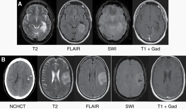
Figure 4.
Imaging characteristics of IDH-mutant gliomas. (A) MRI features of astrocytoma, IDH-mutant, grade 2. This left posterior temporal lobe tumor demonstrates hyperintensity on T2-weighted and fluid-attenuated inverted recovery (FLAIR) sequences. Note the decreased signal within the core of the tumor on FLAIR compared to T2 (T2/FLAIR mismatch). The tumor does not demonstrate decreased signal on susceptibility-weighted imaging (SWI) or contrast enhancement after administration of Gadolinium (Gad). (B) MRI features of oligodendroglioma, IDH-mutant, 1p/19q codeleted, grade 2. This left frontal tumor demonstrates hyperdensities on non-contrast head CT (NHCHT), consistent with calcifications (also seen on SWI). The tumor is hyperintense on T2 and FLAIR and does not demonstrate contrast enhancement after administration of Gad.

