Figure 7.
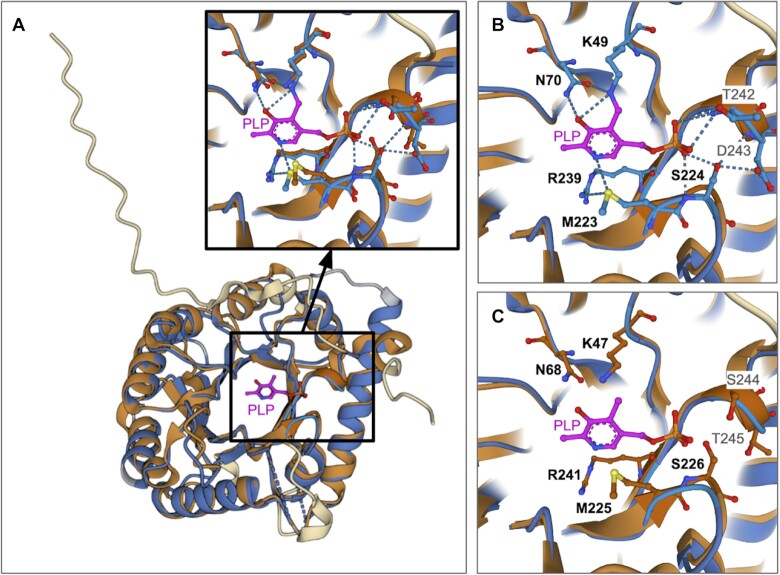
Pairwise superposition of CSM ID AF_AFO94903F1 and PDB ID 1b54. (A) Pair of aligned structures, with both polypeptide chains rendered using ribbon representations. Aligned portions of the PDB structure and CSM are color-coded blue and brown, respectively. Dashed blue lines represent parts of the polypeptide chain not resolved in the X-ray crystallographic experiment. Portions of the PDB structure and CSM that could not be aligned are color-coded gray and cream, respectively. PLP is shown in magenta ball-and-stick, and water molecules are shown as gray spheres. Inset is a closeup of the amino acid residues within 5 Å of the ligand in both the PDB structure and CSM. (B) Same view as in A-inset but showing the amino acid side chains from PDB ID 1b54 that interact with PLP. (C) Same view as in A-inset but showing amino acids from the CSM corresponding to the residues shown in panel (B). Conserved amino acids shown in panels (B) and (C) are identified in bold font. Atom colored coding: C-light blue, brown or magenta; N-dark blue; O-red; S-yellow. Dotted blue lines denote hydrogen bonds and charge–dipole interactions.

