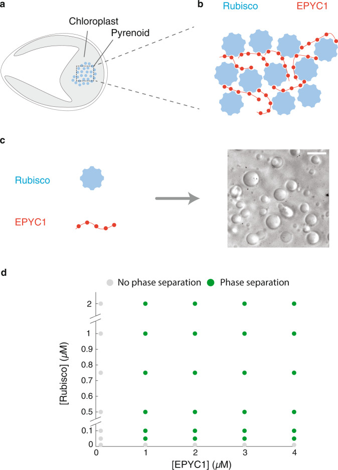
Fig. 1. The components of the pyrenoid—EPYC1 and Rubisco—phase separate in vitro over a broad range of protein concentrations.
a Sketch of a Chlamydomonas reinhardtii cell, highlighting the chloroplast and the pyrenoid. The blue circles indicate Rubisco holoenzymes in the pyrenoid matrix. b Cartoon illustrating that the pyrenoid matrix is held together by multivalent interactions between EPYC1 and Rubisco. c Purified EPYC1 and Rubisco, when mixed together, phase separate in vitro. Scale bar, 5 μm. d Phase diagram of Rubisco-EPYC1 phase separation. Concentrations are expressed in terms of Rubisco holoenzymes and EPYC1 proteins.

