Fig. 1.


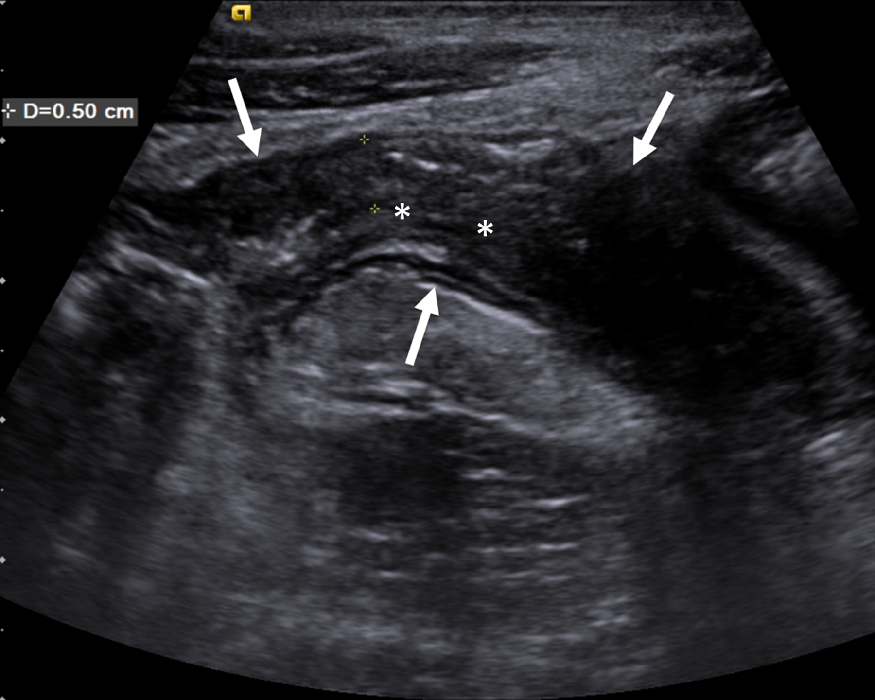
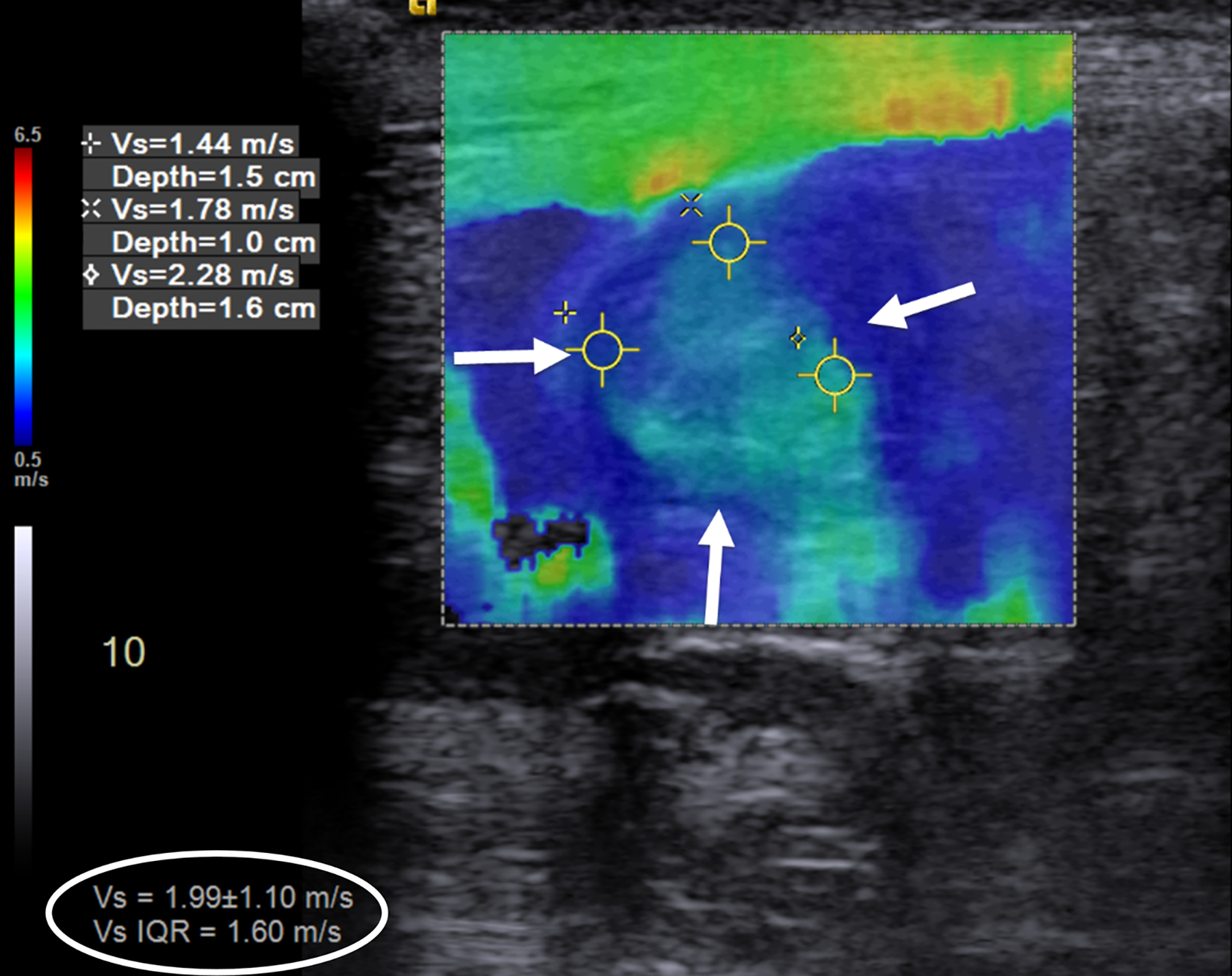
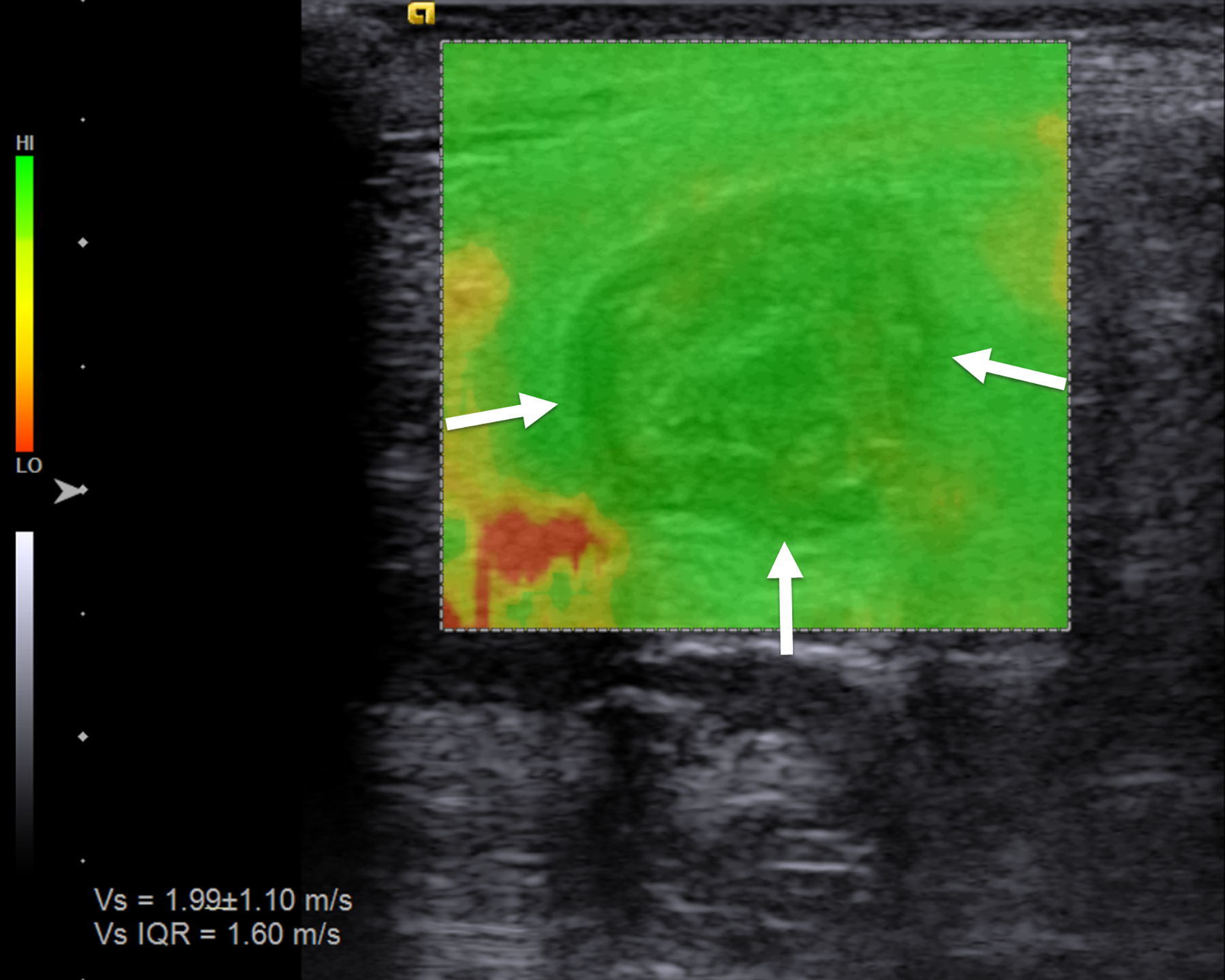
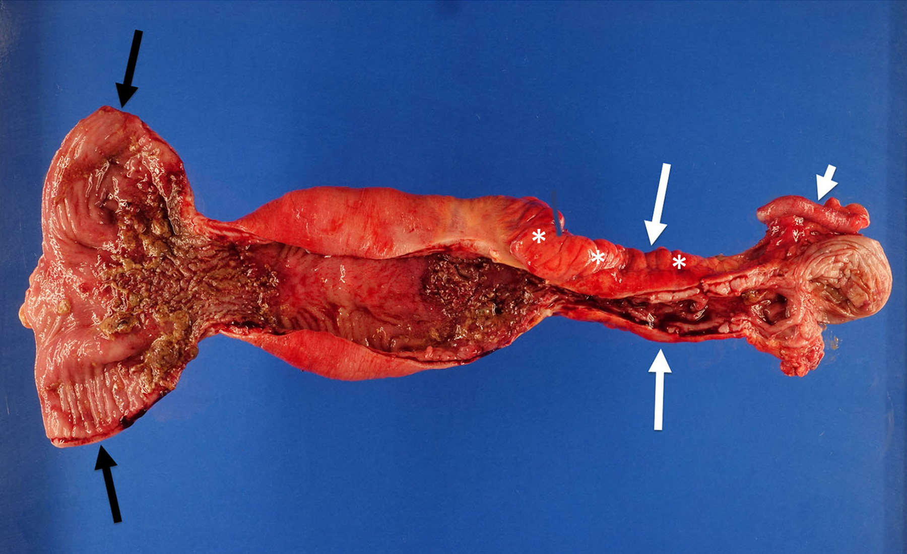
An 18-year-old man with stricturing Crohn disease affecting the terminal ileum. a A longitudinal gray-scale ultrasound image of the terminal ileum (arrows) shows wall thickening and mural heterogeneity, with loss of expected layering. The lumen is narrowed due to a stricture, completely effaced at the level of the asterisks. b A transverse two-dimensional ultrasound shear wave elastography image of the terminal ileum through the mid stricture (arrows) with 10% abdominal strain shows heterogeneous bowel wall stiffening, ranging from 1.44 to 2.28 m/s based on regions of interest placed at the 9:00, 12:00 and 3:00 o’clock locations (yellow symbols). Overall right lower quadrant median shear wave speed within the elastogram is 1.99 m/s (measurement in white oval). c A correlative elastogram quality map confirms that measurements in the bowel wall (arrows) are of “high” quality with regard to shear wave tracking and shear wave speed measurements (based on green color). d A gross pathology image of the resected bowel shows a stricture of the terminal ileum with luminal narrowing (long white arrows) and fat wrapping (so-called creeping fat, asterisks). The uninvolved proximal margin (black arrows) and appendix (short white arrow) are visible
