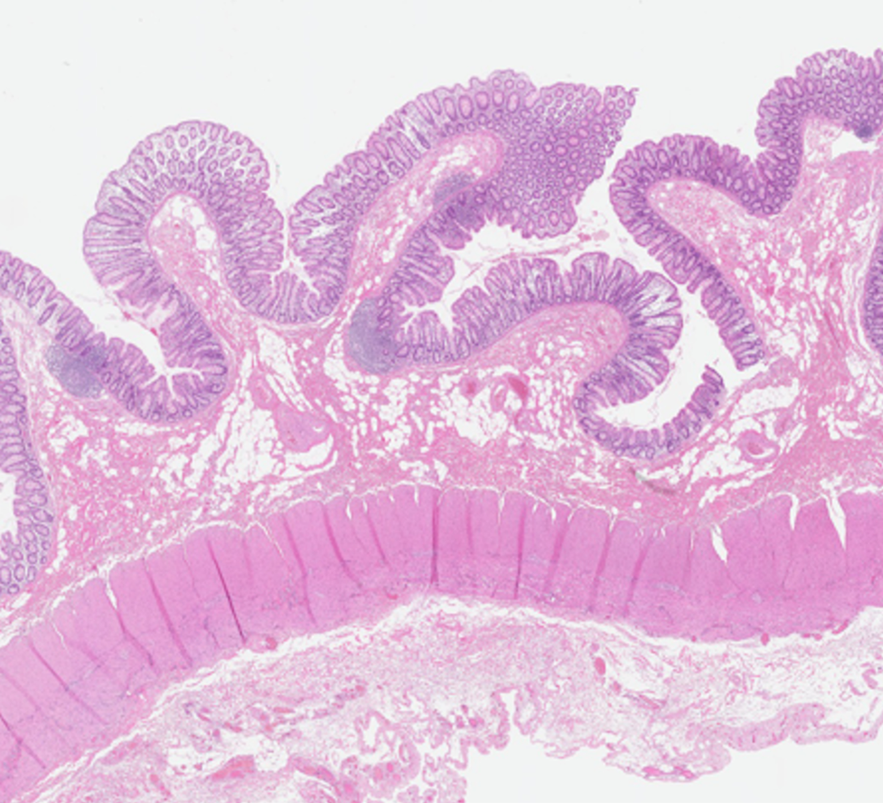
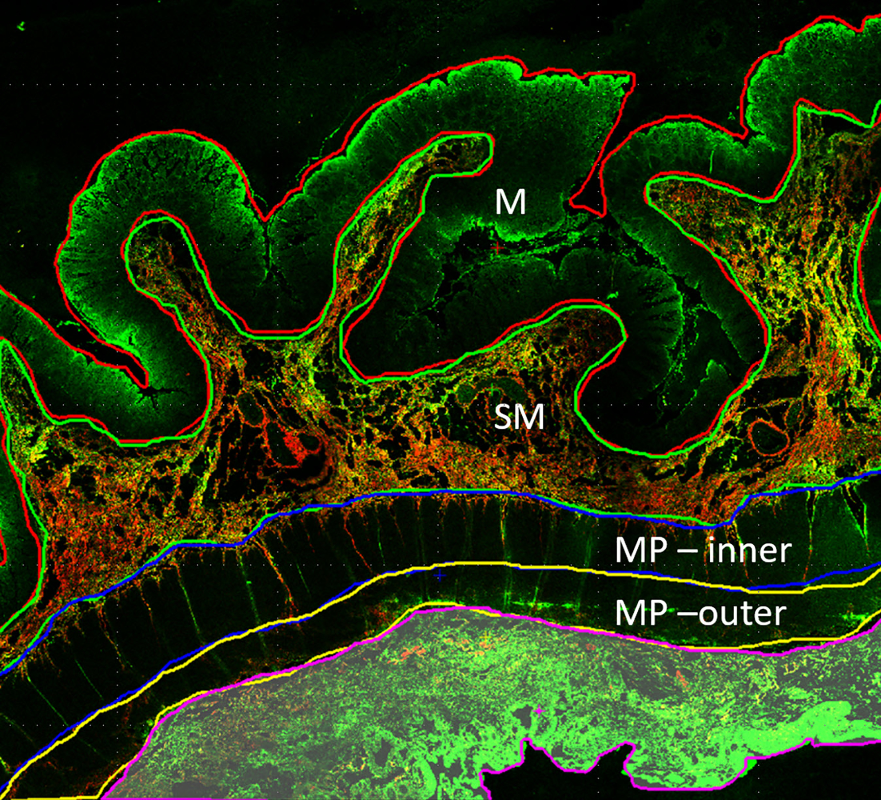
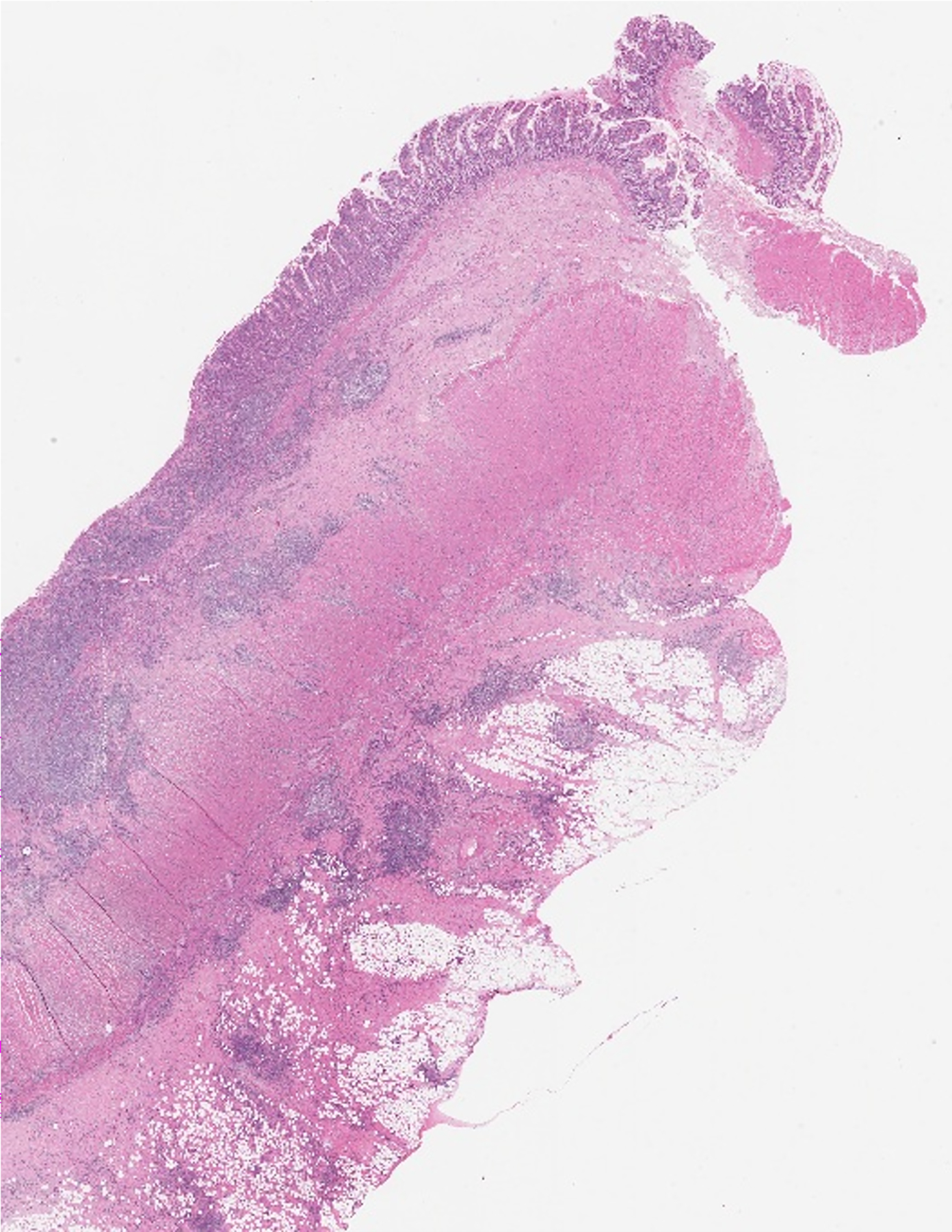
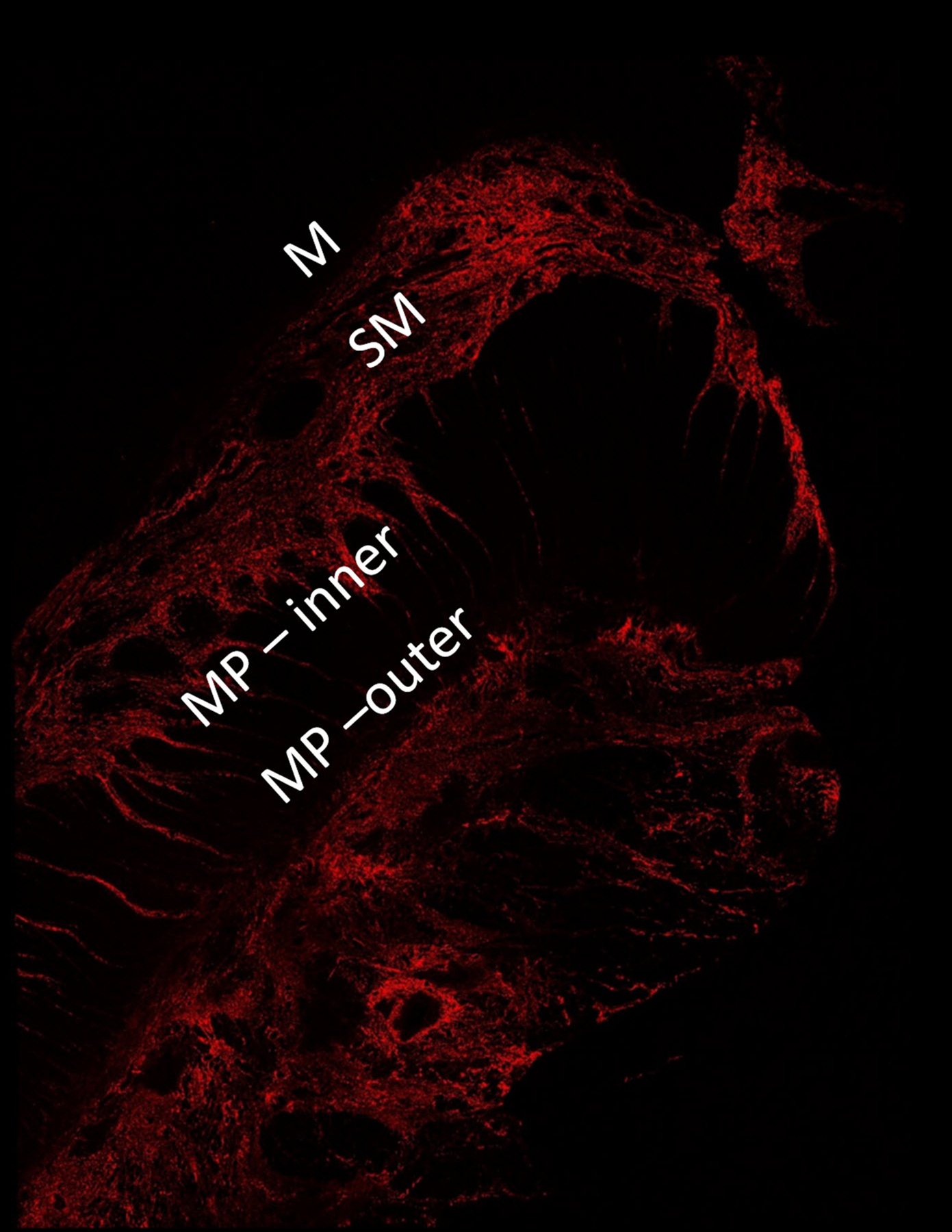
Fig. 2.


A 16-year-old girl with stricturing Crohn disease affecting the terminal ileum. Hematoxylin and eosin stained transmural bowel wall histological specimens from the (a) unaffected small bowel surgical resection margin and (c) mid stricture, respectively. The unaffected margin appears normal, while the stricture shows abundant lymphoid tissue in the mucosa, submucosa and serosa as well as thickening of the muscularis propria. Confocal microscopy images from the (b) surgical margin and (d) stricture. Second harmonic imaging microscopy collagen signal is color-coded in red, while the non-collagen signal is color-coded in green. Colored lines separating the various layers of the bowel wall in figure part (b) were manually drawn using NIS-Elements imaging software (Nikon Instruments Inc., Melville, NY). Only collagen signal from the bowel wall is presented in figure part (d). M mucosa, SM submucosa, MP–inner muscularis propria, inner (circular) layer, MP – outer muscularis propria, outer (longitudinal) layer
