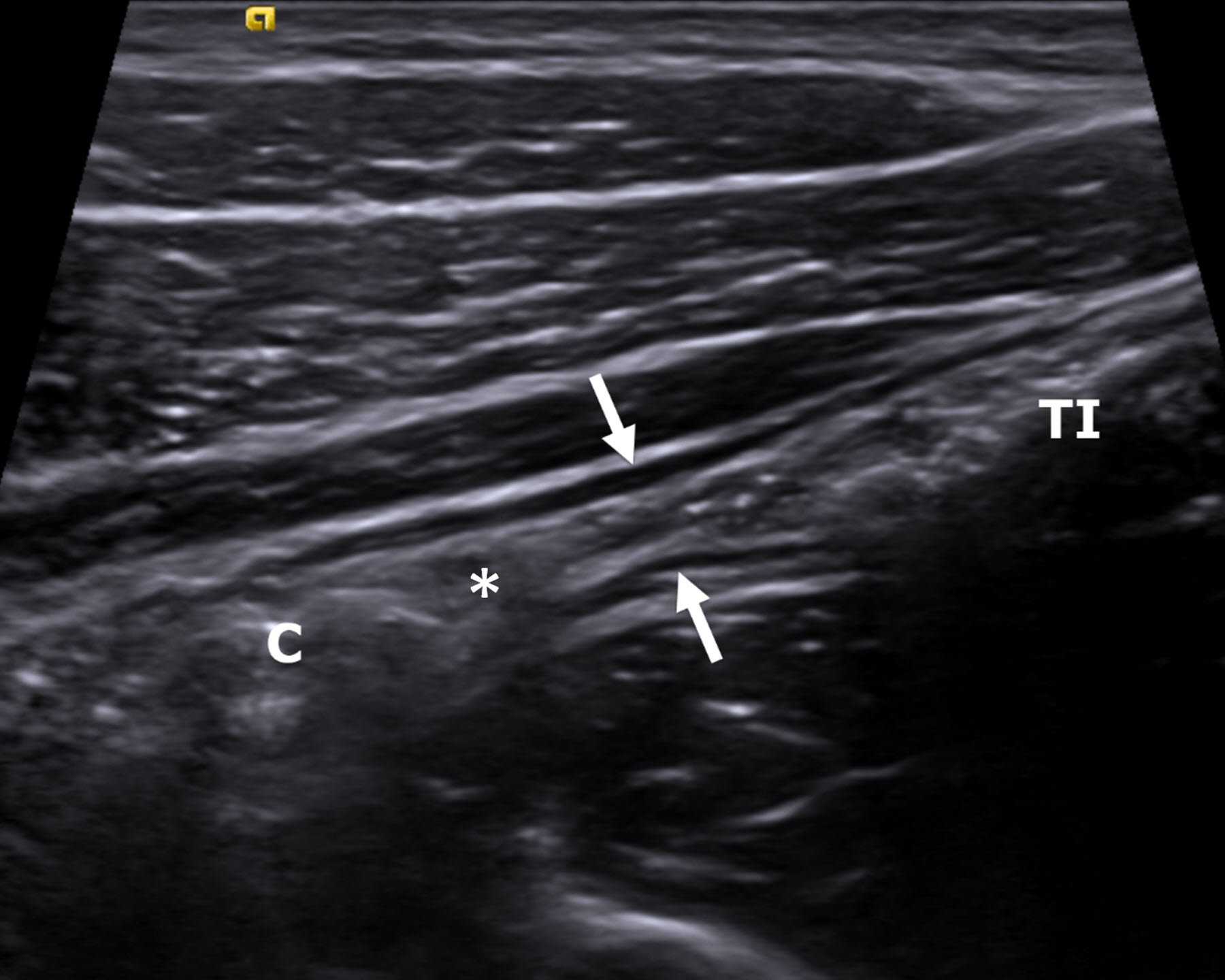
Fig. 3.

An 18-year-old man with Crohn disease and stricture based on the inability to pass an endoscope through the ileocecal valve. A longitudinal gray-scale ultrasound image of the terminal ileum (between arrows) shows luminal narrowing in the absence of bowel wall thickening. There is prominence of the upstream terminal ileum (TI) with intraluminal debris (small bowel feces). The ileocecal valve (*) and colon (C) are visible. Ultrasound shear wave elastography was not possible in this participant, as the bowel wall was too thin to reliably measure shear wave speed
