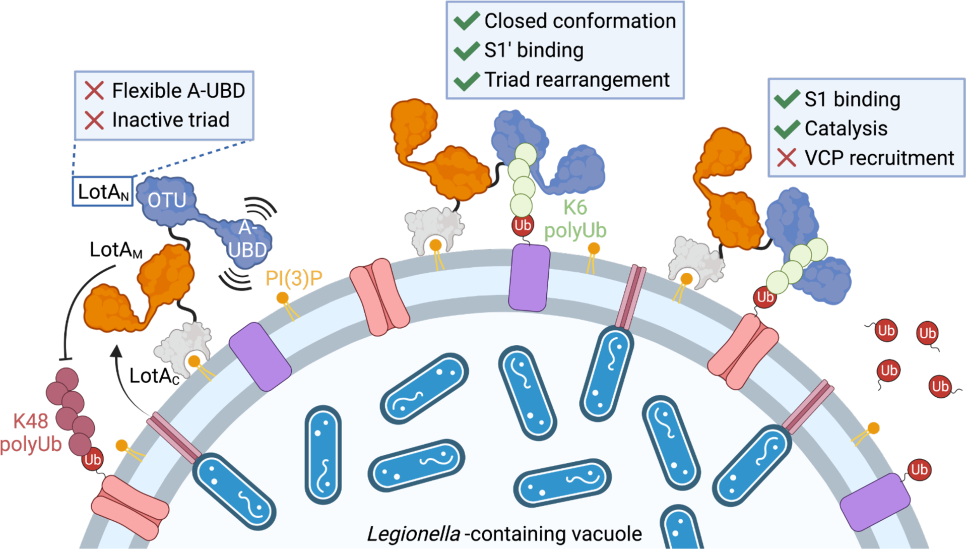
Figure 7: Model for LotA deubiquitinase activity.

Secreted LotA localizes back to the cytosolic face of the LCV via the PI(3)P-binding LotAC domain. At the LCV, DUB activity of LotAM restricts long K48 and K63 polyUb. LotAN is kept inactive by a flexible A-UBD and inactive arrangement of the catalytic triad. Occupying a closed OTU:A-UBD conformation allows formation of an S1’ Ub-binding site. Binding of a K6-linked Ub into the S1’ site orients the LotAN catalytic triad, allowing for hydrolysis of K6 polyUb and prevention of VCP recruitment. Created with BioRender.com.
