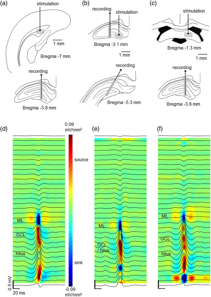
FIGURE 1.

The effect of dentate spikes on responses to electrical stimulation was studied in the entorhinal cortex to the dentate gyrus (EC‐DG) synapse (a), the DG‐CA3 synapse (b) and the CA3‐CA1 synapse (c) in adult male Sprague–Dawley rats under urethane anesthesia. Approximate stimulation and recording sites are illustrated with gray shading. (d, e, and f) panels illustrate the current source density map (blue = sink, red = source) and average local‐field potential traces during dentate spikes recorded from representative rats in each respective experiment. Note that the data for (d–f) are from the 1‐h baseline recording during which no stimulations were administered. A sink is visible in the molecular layer and source in the DG during dentate spikes
