
Figure 2.
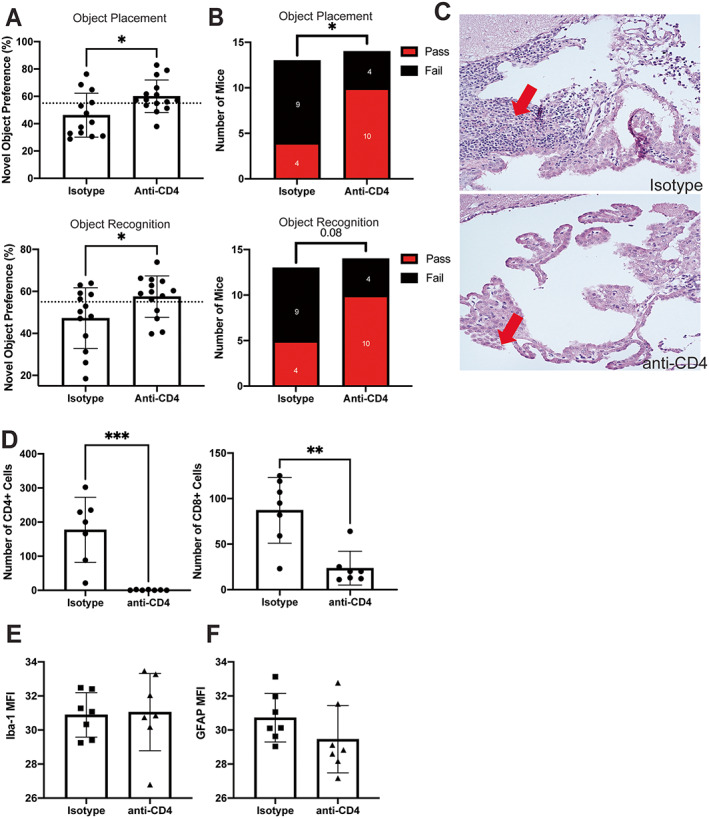
Improved cognition in MRL/lpr mice after systemic depletion of CD4+ T cells. A, Percentage of mice exhibiting novel object preference in assessments of spatial memory (object placement test) and recognition memory (object recognition test). Dashed line indicates a cutoff level of 55% for pass/fail, where those above the line had a passing score (i.e., showed novel object preference). B, Number of mice with a passing or failing score, based on 55% cutoff, during novel object preference tests for either spatial memory (n = 13 mice) (top) or recognition memory (n = 14 mice) (bottom). C, Representative images of hematoxylin and eosin–stained brain tissue from mice treated with isotype control or anti‐CD4, with arrows indicating the choroid plexus (CP) infiltrate. D–F, Immunofluorescence staining (expressed as mean fluorescence intensity [MFI]) of CD4+ and CD8+ in T cells in the CP (D), of ionized calcium–binding adapter molecule 1 (IBA‐1) in the hippocampus to assess microglial activation (n = 7 mice) (E), and of glial fibrillary acidic protein (GFAP) in the hippocampus to assess astrocyte activation (F). In A and D–F, solid symbols show individual mice; bars show the mean ± SD. The treatment groups were compared using Student's t‐test or chi‐square test. * = P < 0.05; ** = P < 0.01; *** = P < 0.001. Color figure can be viewed in the online issue, which is available at http://onlinelibrary.wiley.com/doi/10.1002/art.42252/abstract.