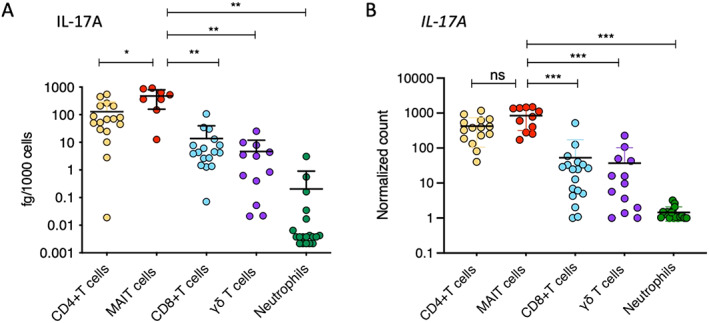
Figure 2.

IL‐17A protein production after 18 hours of stimulation (A) and IL17A transcript levels (normalized expression) after 2 hours of stimulation (B) were assessed in sorted CD4+ T cells, mucosal‐associated invariant T (MAIT) cells, CD8+ T cells, γδ T cells, and neutrophils from the peripheral blood of axial SpA patients. Cells were stimulated with phorbol myristate acetate (50 ng/ml), calcium ionophore A23187 (5 μM), and β‐1,3‐glucan (50 μg/ml). Symbols represent individual samples. Bars show the mean ± SEM. * = P < 0.05; ** = P < 0.01, by Mann‐Whitney test. *** = P < 0.001, by Wilcoxon‐Mann‐Whitney test. ns = not significant (see Figure 1 for other definitions).
