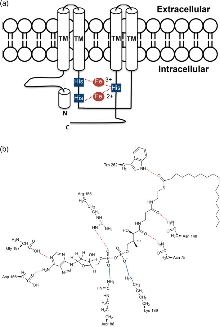
FIGURE 6.

Panel (a) is a schematic representation of the topological model of the acyl‐CoA membrane‐bound desaturases. The white cylinders represent the transmembrane domains (TM). The small blue squares highlight the conserved histidine boxes, that coordinate to the di‐iron centre and determine the correct orientation of the protein in the membrane and interactions with the substrate in a selective and stereospecific manner. Panel (b) shows the interactions between the amino acid residues of the active site and the stearoyl‐CoA substrate. The red dotted lines highlight the H‐bonds. The blue lines highlight the electrostatic interactions. 55
