
Figure 2.
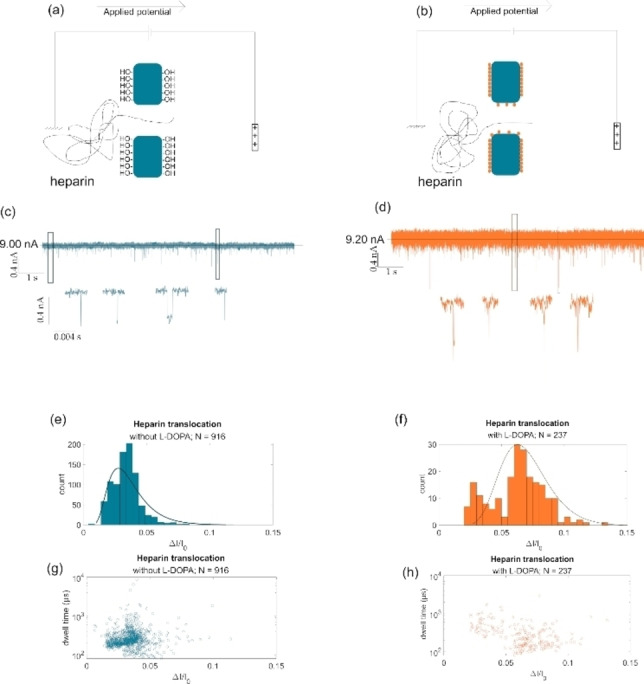
Sketch of heparin translocation in (a) raw SiN and (b) L‐dopa coated SiN nanopores under +300 mV in 2 M NaCl PBS solution. 10 seconds sample of the current trace for heparin translocation in (c) uncoated and (d) coated SiN nanopore. The full current trace is 5 min long and generally have a baseline of 9 n. Current blockades in the form of histograms for heparin translocation in (e) uncoated (N=916) and (f) coated SiN (N=237) nanopore. Scatter plots showing the translocation events in dwell time (μs) in logarithmic scale versus the current blockade (ΔI/I0) for heparin translocation in (g) uncoated and (h) coated SiN nanopore.