
Figure 5.
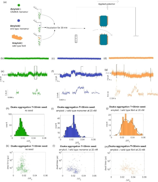
(a) Sketch of Aβ(42) Osaka aggregate preparation and detection through an L‐dopa coated SiN nanopores Current trace at t=0 min taken at nanopore 10 nm in diameter in 3 M LiCl+4 mM HEPES at −300 mV for Aβ(42)‐E22Δ (b), with Aβ(42)‐WT monomers (c), and with 20 nM Aβ(42)‐WT fibrils (d); current trace at t=30 min taken at nanopore 10 nm in diameter in 3 M LiCl+4 mM HEPES at −300 mV for Aβ(42)‐E22Δ variant (e), with Aβ(42)‐WT monomers (f), and with 20 nM Aβ(42)‐WT fibrils (g) generally the baseline measured was around 10 nA; Histogram showing the registered ΔI/I at T=30 min in 5 min of measurement for Aβ(42)‐E22Δ (N=344) (h), with Aβ(42)‐WT monomer (N=244) (i), and 20 nM Aβ(42)‐WT fibril (N=164) (j); scatter plot showing ΔI/I against Δt (μs) in logarithmic scale for these same events for Aβ(42)‐E22Δ (k), with Aβ(42)‐WT monomer (l), and with 20 nM Aβ(42)‐WT fibril (m).