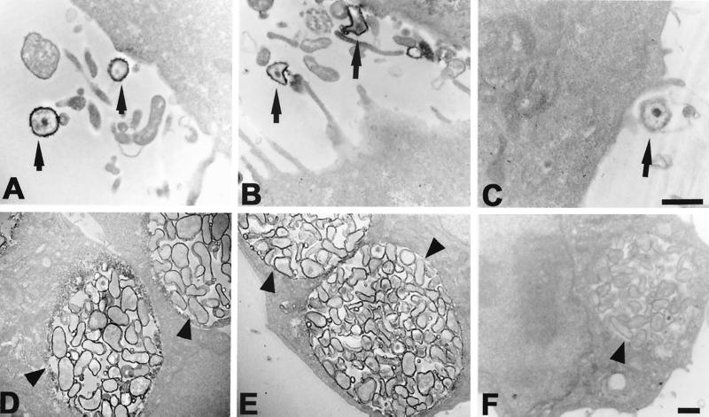
FIG. 2.
Transmission electron microscopy of C. pneumoniae EBs (A) and RBs (D) stained with the GZD1E8 MAb indicate surface localization of the antigenic determinant. Surface staining of C. pneumoniae EBs (B) and RBs (E) is also shown with an anti-chlamydial LPS MAb, EVIH1. No staining of C. pneumoniae EBs (C) and RBs (F) was observed with anti-rickettsial MAb 8-13A4A10. Arrowheads indicate inclusions and arrows indicate EBs of C. pneumoniae. Bars = 0.5 μm.

