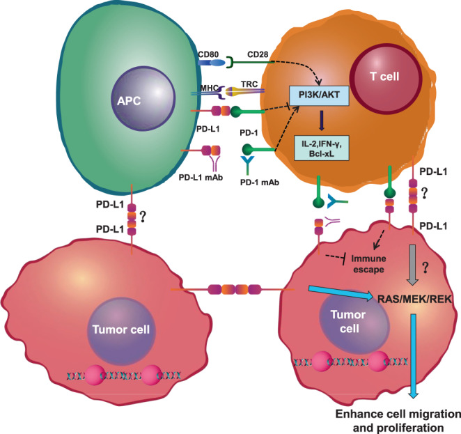
FIGURE 8.

Schematic of PD‐L1 interaction with PD‐L1. PD‐1 from T cells binding to PD‐L1 on antigen‐presenting cells inhibits the PI3K/AKT pathway. PD‐1 ultimately decreases the induction of cytokines, such as IFN‐γ, and cell survival proteins, such as Bcl‐xL. T cells can recognize their target antigen peptide/MHC presented on tumour cells and initiate tumour‐cell lysis. Tumour cells can express PD‐L1, which binds to PD‐L1. APC, antigen‐presenting cell; IL‐2, interleukin 2; MHC, major histocompatibility complex; PD‐1, programmed cell‐death protein 1; PD‐L1, programmed cell death 1 ligand 1; TCR, T‐cell receptor
