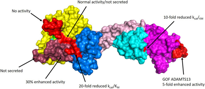
FIGURE 3.

ADAMTS13 exosites. The crystal structure of MDTCS is shown, with domains indicated: metalloprotease (yellow), disintegrin‐like (blue), TSP1 (rose), cysteine‐rich (cyan), and spacer (magenta). The active site zinc (cyan) and catalytic glutamic acid (mutated to glutamine; green) are also indicated within the metalloprotease domain. The structure was obtained using a stabilizing antibody fragment to the metalloprotease domain (not shown). Compared with the AlphaFOLD2 model, this structure is lacking two loops in the cysteine‐rich domain that did not resolve (Asp453‐Tyr468 and I490‐K497). A summary of important mutagenesis work is indicated, with modified residues indicated in light red and dark red for clarity. These studies highlight important contact points for the VWF A2 domain to exosites on ADAMTS13. (PDB: 6qig)
