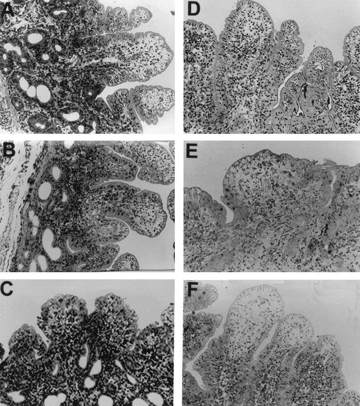
FIG. 7.
Cross sections of intestinal mucosa from ovine ileal loops stained with hematoxylin and eosin and incubated for 3 h with Salmonella serovars. (A) Untreated control; intestinal mucosa infected with (B) serovars Abortusovis (strain SS44), (C) Dublin (strain SD2229), (D) Gallinarum (strain G9), (E) Typhimurium strain 4/74, and (F) Typhimurium strain 4/74 InvH. A minimum of 10 sections were examined for each loop infected (n = 6) with each Salmonella strain. Representative sections are shown. Magnification, ×400.

