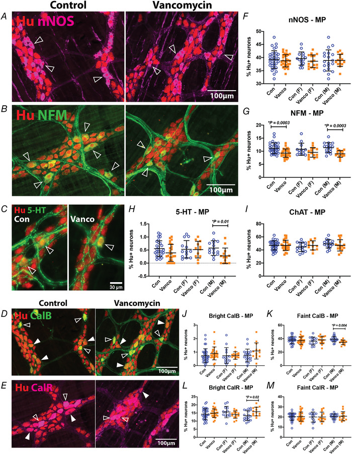
Figure 3. Neonatal exposure to vancomycin had sexually dimorphic effects on the neurochemistry of myenteric neurons.

A–E, representative images of myenteric plexus preparations from the mid colon of control‐ and vancomycin‐treated 6‐week‐old mice. A, myenteric neurons were stained with the pan‐neuronal marker, Hu (red) and nNOS (magenta). Some nNOS‐immunoreactive Hu+ neurons are indicated (open arrowheads). Myenteric neurons were stained with Hu (red) and NFM (B), 5‐HT (C) or calbindin (D) in green. Some NFM−, 5‐HT‐ or calbindin‐immunoreactive Hu+ neurons are indicated (open arrowheads). The calbindin antiserum stains some neurons brightly (open arrowheads) and others faintly (closed arrowhead). E, myenteric neurons were stained with Hu (red) and calretinin (magenta). The calretinin antiserum stains some neurons brightly (open arrowheads) and others faintly (closed arrowhead). F–M, quantification of nNOS+ (F), NFM+ (G), 5‐HT+ (H), ChAT+ (I), bright calbindin+ (J), faint calbindin+ (K), bright calretinin+ (L) and faint calretinin+ (M) neurons relative to Hu+ neurons in the mid colon. For Hu/nNOS: Control female (n = 15), Vanco female (n = 16), Control male (n = 19), Vanco male (n = 16). For Hu/NFM: Control female (n = 13), Vanco female (n = 15), Control male (n = 17), Vanco male (n = 15). For Hu/5‐HT: Control female (n = 13), Vanco female (n = 12), Control male (n = 14), Vanco male (n = 14). For Hu/ChAT: Control female (n = 15), Vanco female (n = 11), Control male (n = 16), Vanco male (n = 15). For Hu/calbindin: Control female (n = 14), Vanco female (n = 15), Control male (n = 19), Vanco male (bright: n = 13, faint n = 12). For Hu/calretinin: Control female (n = 13), Vanco female (n = 14), Control male (n = 18), Vanco male (n = 13). Data represented as means ± SD. Treatment groups were compared statistically using unpaired t‐tests or the Kolomogorov–Smirnov test (for panel H specifically).
