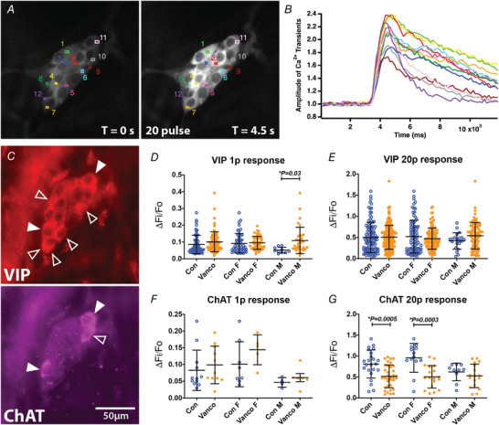
Figure 5. Neonatal exposure to vancomycin affected the amplitude of electrically evoked [Ca2+]i transients in submucosal plexi.

A, regions of interest (ROI 1–12) drawn on representative fluorescence micrographs of the 20 pulse train (20 Hz)‐evoked [Ca2+]i response in submucosal neurons of 6‐week‐old mice given control‐treatments as neonates (GCaMP3 signal at rest (t = 0); and during 20 pulse train stimulation, T = 4.5 s). B, amplitude of [Ca2+]i transients from cells stimulated with a 20 pulse train stimulation. Colours correspond to the ROIs in A. C, post hoc immunohistochemistry labelling vasoactive intestinal peptide (VIP) and choline acetyltransferase (ChAT). Open arrowheads indicate neurons positive for VIP or ChAT. Filled arrowheads indicate neurons positive for both VIP and ChAT. D and E, amplitude of [Ca2+]i transients (ΔF i/F 0) from VIP cells stimulated with a single stimulus (D) and a 20 pulse train (E). F and G, amplitude of [Ca2+]i transients (ΔF i/F 0) from ChAT cells stimulated with a single stimulus (F) and a 20 pulse train (G). All graphs show means ± SD. Treatment effects were assessed with a two‐tailed unpaired t‐test.
