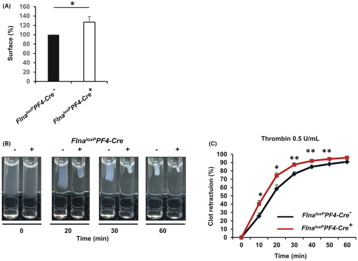
FIGURE 6.

Outside‐in signaling of washed platelets. A, Platelet spreading was performed on a fibrinogen matrix (100 μg/ml). Platelets were incubated at 37°C for 30 min and then stained with Alexa Fluor488‐labeled phalloidin (0.3 μM). Platelets were visualized under an epifluorescence microscope (Nikon, Eclipse 600). Cell surfaces were analyzed using the Fiji software. The graph represents the means ± standard error of the mean (SEM) of four independent determinations. *p < .05, (unpaired Student t‐test). B, Clot retraction of washed platelets was initiated by adding fibrinogen (500 μg/ml) and thrombin (0.5 U/ml). Photographs were taken at different times and (C) the area of clot was measured using the Fiji software. The graph represents the means ± SEM of four independent determinations. *p < .05, **p < .01 (unpaired Student t‐test)
